
Application Area
Immune System

Groundbreaking developments in molecular genetics and cellular electrophysiology have provided newfound knowledge on the crucial role that ion channels play in the immune system. The advent and adoption of automated patch clamp systems have allowed researchers to investigate ion channels at an exponentially faster pace than manual patch clamp.
The implications of ion channels in various immunological conditions and their modulation by autoimmune diseases cannot be overstated.
Autoimmune channelopathies
- Lambert-Eaton syndrome (LEMS)
- Isaacs’ syndrome
- Myasthenia gravis (MG)
- Chronic kidney disease (CKD)
Further exploration of the role of ion channels may lead to innovative therapeutic interventions that can significantly improve the lives of individuals affected by these conditions.
Below, we highlight key ligand and voltage-gated ion channels that have been shown to play a significant role in the immune system.
IonFlux Mercury 16 is especially suited for research into immune cell ligand-gated and mechano-sensitive ion channels. Ion channels can be transfected into recombinant cell lines or induced pluripotent stem cells (iPSC).
Important System Characteristics:
- Fast solution exchange
- Continuous flow
- Temperature control
Nicotinic Acetylcholine Receptors
Nicotinic Acetylcholine Receptors (nAChRs) are not only expressed in the peripheral and central nervous systems but also in the immune system. Research has shown a novel role for the α7nAChR in mediating macrophage recruitment to inflamed tissue. In a more recent publication, electrophysiological recordings using IonFlux from primary monocytes showed the clear expression of α7 activated by 60 mM ACH and enhanced by PNU120596.
GABA Receptors
GABA A Receptors are well known in the central nervous system, however, it is also established that they play a significant role in the immune system. They are found in various immune cells, including mononuclear phagocytes and lymphocytes. They affect cell migration, cytokine section and immune cell activation among other functions.
Mechanosensitive ion channels
Piezo1 and Piezo2 ion channels convert external mechanical forces (such as stretching, pressure, and shear stressThe tangential force per unit area from flowing fluid; a key parameter in vascular biology, platelet function, leukocyte rolling, and biofilm adhesion studies.) into electrical and chemical signals. Activation of these channels plays a vital role in chronic inflammatory diseases. Pharmacological targeting of Piezo1 could be a promising approach in managing inflammatory conditions.

Transient Receptor Potential
TRPV4 (Transient receptor potential vanilloid 4) is a calcium-permeable ion channel expressed in multiple cells including immune cells. It responds to mechanical forces and contributes to immune cell activation and inflammation. It significantly influences macrophage behavior in the lungs, influencing migration, phagocytosis, and cytokine secretions. Other important channels are TRPM2. TRPM4 and TRPM7.
Glutamate Receptors
Although primarily associated with neurons, NMDA receptors are also expressed in immune cells. Recent research indicates the presence of NMDA-type glutamate receptors in cells such as thymocytes, lymphocytes, and neutrophils. The activation of NMDA receptors in lymphocytes leads to pro-inflammatory cytokines such as interleukin 1β, interleukin 6 and tumor necrosis-alpha (TNF-α).
P2X Receptors
P2X receptors are cation-permeable ligand-gated ion channels. They open in response to the binding of extracellular adenosine 5’-triphosphate (ATP). These receptors belong to the ENaC/P2X superfamily and share structural similarities with ENaC channels. P2X7 causes calcium influx in T cells. Other than regulating T cell activation, in the presence of high levels of extracellular ATP, the activity of P2X7 receptors can lead to cellular apoptosis.
Representative Channels
- nAChR
- GABA(A) R
- TRPV4, TRPM(2,4,7)
- NMDAR
- PIEZO1
- P2X7
Potassium Channels
Kv1.3 Channels are present predominantly in T-Lymphocytes. These delayed rectifier K channels facilitate the calcium influx necessary for lymphocyte activation and proliferation. They generate potassium diffusion potential across plasma membranes, regulating resting membrane potential and controlling cell volume. The rise in intracellular calcium concentration activates calcineurin, which dephosphorylates the nuclear factor of activated T cells (NFAT). NFAT then accumulates in the nucleus and binds to the promoter of the gene encoding interleukin 2 (IL-2), a critical cytokine for immune response. Some commonly used drugs, such as nonsteroidal anti-inflammatory drugs (NSAIDs), macrolide antibiotics, and calcium channel blockers (CCBs), can suppress Kv1.3 channel currents in lymphocytes. Consequently, they exert immunosuppressive effects.
KCa3.1 is a second type of K+ channel that is expressed in T cells. This channel is not sensitive to voltage but to the rise to cytosolic calcium. Activation of these channels also leads to efflux of potassium helping in repolarization of the membrane. KCa3.1 is closed under resting conditions but opens rapidly if the intracellular Ca2+ concentration rises.
Calcium Channels
Voltage gated calcium channels play an important role in T Cells. Both the L-type and the T-Type are expressed and are involved in T cell receptor (TCR) mediated calcium signaling. Recently Cavβ1 has been shown to be involved in cell expansion and apoptosis. Other Calcium channels like CRAC (calcium release activated channels) are activated by calcium release from the endoplasmic reticulum play also play an important role in T Cell activities.
Representative Channels
- KV1.3
- KCA3.1
- CRAC

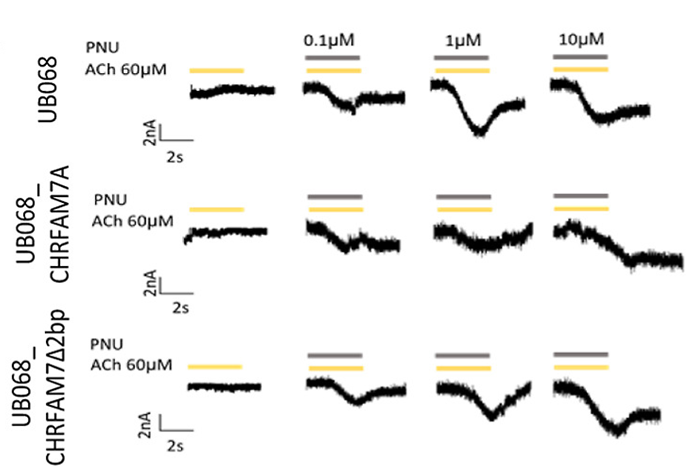
Article Highlight
CHRFAM7A diversifies human immune adaption through Ca2+ signaling and actin cytoskeleton reorganization
In this newly published article, Szigeti et al. set out to better understand immune function and reduce the translational gap between rodent and human studies by altering CHRFAM7A, a unique human fusion gene between CHRNA7 and ULK4. Human induced pluripotent stem cells (iPSC) were differentiated into mature macrophages and an IonFlux 16 automated patch clamp system was used to assess Ca2+ influx in response to the acetylcholine and a positive allosteric modulator of α7-nAChR, PNU-120596.
Customer Spotlight
Predicting Unwanted Drug/Heart Interactions with Artificial Intelligence
