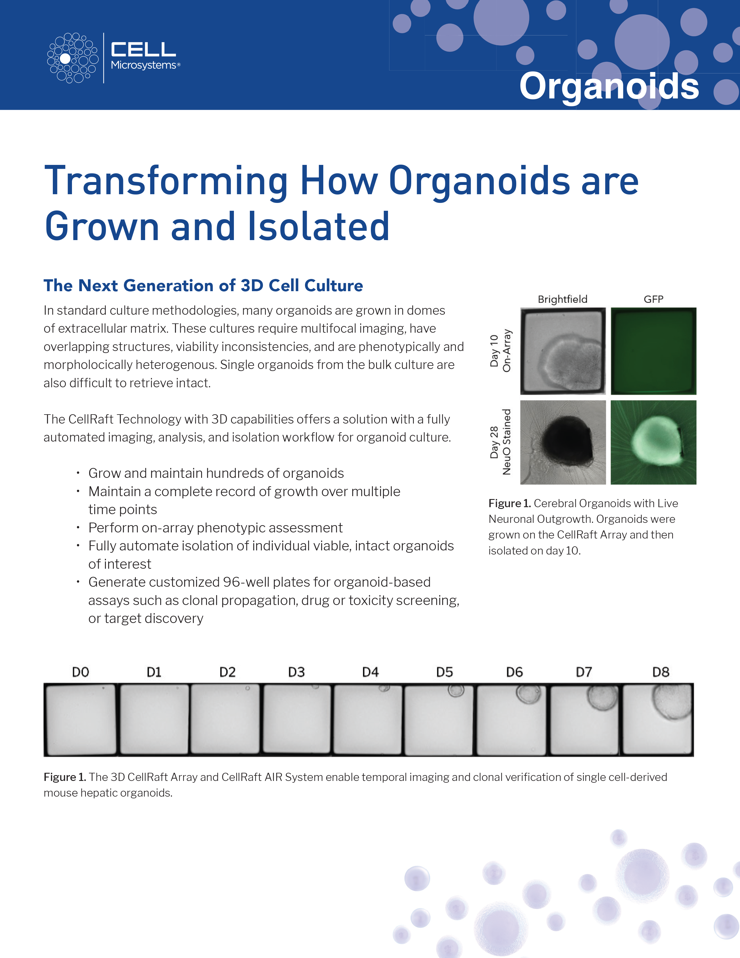
In typical organoid cultures, the structures overlap, requiring multifocal imaging with expensive equipment. They grow at different sizes and rates, leading to heterogeneity in viability and phenotype, resulting in confusing and uninterpretable data. Organoids grown in bulk culture are stuck in viscous extracellular matrix and are difficult to retrieve intact.
The CellRaft AIR® System with 3D capabilities offers a solution with a fully automated imaging and isolation workflow for organoid culture.
Grow and maintain hundreds of organoids
Maintain a complete record of growth over multiple time points
Perform on-array phenotypic assessment
Fully automate isolation of individual viable, intact organoids of interest
Generate customized 96-well plates for organoid-based assays such as clonal propagation, drug or toxicity screening, or target discovery
Capture images in brightfield and fluorescence throughout the full height of the organoid with z-stack imaging.
The images showing detailed phenotypic characterization are cataloged and easily exported for the desired use.
Our user-friendly proprietary software enables a one-touch full-array image scan
Brightfield Imaging of Mouse Hepatic Organoids
EpCAM & Hoechst Imaging of Mouse Hepatic Organoids
Identify
Identify CellRafts containing 3D structures of interest using CellRaft Cytometry™ to detect organoids. Visually verify single cell clonality, morphological data, and growth rate, critical data points for workflows requiring track and traceability to confirm clonality. Identified organoids can be further evaluated and selected for phenotypic characteristics, such as organoid diameter, circularity, and fluorescence intensity.
Perform phenotypic characterization with Raft Cytometry.
Identify and verify CellRafts containing 3D structures of interest and create sets of sub-populations for isolation
Isolate
The Air System with 3D capabilities enables automated isolation of live, intact organoids without fixation, dissociation, or damage to the 3D structure. Organoids remain viable through isolation and continue to grow off-array.