Leukocyte Adhesion Cascade
Study all steps of the leukocyte adhesion cascade with one system
The leukocyte adhesion cascadeA multistep process that facilitates the recruitment and migration of white blood cells to sites of inflammation or infection; observed in several steps, rolling adhesion, firm adhesion, transmigration. consists of capture, rolling, slow rolling, arrest, strengthening/spreading, and transmigration. Typically, analyses of these steps require at least 3 separate systems and/or assays.
BioFlux shear flow systems enable the visualization and analysis of all of the steps of the leukocyte adhesion cascade under physiological/pathological shear flowFluid movement that applies force to cells and biofilms; used to mimic physiological/pathological conditions. using one system.
General BioFlux workflow for cell adhesion cascade assays

Leukocyte adhesion cascade assays using BioFlux
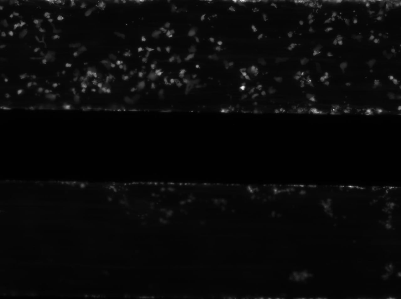
Adhesion: Recombinant human vimentin binds to P-selectin and blocks neutrophil capture and rolling on platelets and endothelium
The top channel shows the inhibition of neutrophil adhesion by recombinant human vimentin. The bottom channel shows neutrophil adhesion under control conditions.
Rolling: The vimentin rod domain blocks P-selectin-P-selectin glycoprotein ligand 1 interactions to attenuate leukocyte adhesion to inflamed endothelium
Neutrophil rolling on endothelial cells under shear flow. Scale bar = 50 µm
Transmigration: Ezrin and Moesin Are Required for Efficient T Cell Adhesion and Homing to Lymphoid Organs
Tracking of the transmigration of CMTMR stained wild-type T cells (red) and CFSE stained ERM-deficient T cells (green) through TNF-α treated 3B-11 endothelial cells.

Key BioFlux Advantages For Cell Adhesion Cascade Studies
- Image and quantify live cell adhesion and rolling in a physiological environment using primary endothelial cells, human peripheral blood mononuclear cells, and more
- Utilize time-lapse microscopy and powerful post-experiment image analysis software to determine rolling distance under flow
- Examine up tp 24 different monolayers at one time to determine IC50 concentrations or screen multiple compounds
- Study truly biological cell adhesion and transmigration in the same assay
FAQs:
Immune Cell Adhesion Assays Using the BioFlux
What is the best in vitro model for studying immune cell adhesion under flow?
The BioFlux System provides a microfluidic platform that replicates physiological shearFlow conditions intended to resemble those in vivo (e.g., vasculature, catheters, mucosal surfaces).shear stressThe tangential force per unit area from flowing fluid; a key parameter in vascular biology, platelet function, leukocyte rolling, and biofilm adhesion studies., making it ideal for modeling immune cell adhesion endothelial monolayers or ECM proteins. It enables real-time observation of immune cell dynamics under controlled flow conditions.
How can I study leukocyte-endothelial interactions using microfluidics?
The BioFlux supports culturing endothelial cells directly in microfluidic channels and perfuse leukocytes or other immune cells under defined shear stress. This setup supports quantitative analysis of leukocyte adhesion in response to inflammatory stimuli.
What microfluidic system supports live-cell imaging of leukocyte adhesion?
The BioFlux integrates with standard microscopy platforms (contrast or fluorescence), enabling high-resolution live-cell imaging of leukocyte behavior.
Can the BioFlux be used to study leukocyte recruitment in inflammation models?
Yes, the BioFlux is widely used in immunology research to study leukocyte recruitment during inflammation. Its ability to simulate vascular flow and endothelial activation makes it ideal for investigating immune cell trafficking in response to cytokines or chemokines.
What are the advantages of using BioFlux over static leukocyte adhesion assays?
The BioFlux replicates shear flow conditions that are essential for accurate modeling of leukocyte behavior. Unlike static assays, it allows for controlled shear stress, real-time imaging, and reproducible quantification of cell adhesion under physiologically relevant conditions.
How does BioFlux support multi-condition leukocyte adhesion experiments?
The BioFlux offers well plate formats with 3, 8, and 24 microfluidic channels, allowing researchers to test multiple experimental conditions. This makes it ideal for comparing cytokine treatments, surface coatings, or leukocyte subtypes under flow.
What substrates can be used in the BioFlux microfluidic channels for leukocyte adhesion studies?
Common substrates include endothelial cell monolayers, ICAM-1, VCAM-1, and selectins. BioFlux microfluidic channels can be coated or seeded with endothelial cells to mimic vascular surfaces, enabling targeted studies of leukocyte adhesion mechanisms.
Is the BioFlux compatible with primary leukocytes or whole blood?
Yes, the BioFlux supports perfusion of primary leukocytes, whole blood, or isolated immune cell populations.