
Seed Cells on CellRaft Array
Seeding cells on the CellRaft array is straightforward and comparable to seeding cells on any standard tissue culture dish. The diluted cell suspension is dispensed into the central reservoir, and cells settle into the microwells by gravity. The CellRaft Arrays are designed to isolate a high number of single cells with the added benefit of shared media in a flask-like environment to increase viability and monoclonal outgrowth.

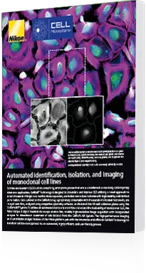
Image Using CellRaft AIR System
Brightfield and fluorescence scanning provides single-cell resolution imaging in as little as seven minutes. CellRafts can be scanned over the days or weeks needed to identify samples of interest without monopolizing the instrument with a single experiment.

Identify Cells of Interest in Software
CellRaft Cytometry™ allows for image-based verification of single cells to ensure monoclonality and analysis of a variety of parameters over time, ranging from morphology to gene expression. Users can easily define the characteristics of the target cells or colonies and map them for software-guided CellRaft selection for downstream isolation.
Automated Isolation to 96-well Plate
After identification, the CellRafts are gently dislodged from the CellRaft Array by a release needle and retrieved from the array by a magnetic wand to transfer the CellRafts into 96-well plates for downstream analysis. The fully automated CellRaft AIR System uses an AI powered, fluidics-free isolation mechanism that incorporates a proprietary glass transfer wand with a surface designed to prevent cross-contamination between wells.

Enhanced Cell Outgrowth Compared to Traditional Methods
The flask-like culture conditions of the CellRaft Array combined with the automated and gentle isolation of identified cells, colonies, or organoids of interest results in a higher outgrowth percentage post-isolation than can be accomplished with limiting dilution or traditional cell sorters and dispensers.


“We recommend the CellRaft Technology for our colleagues struggling with single cell isolation. Even with help from FACS, although single cell clones could be obtained, from our experiences, CellRaft not only speeds up this process by providing many more clones but more importantly, the unique monitoring process provided by CellRaft supplies additional information for every single clone with a higher quality of clones.”

“We can now easily identify thousands of single organoids with various morphologies (size, shape, complexity) and isolate hundreds in less than an hour. We also use the CellRaft AIR System for cloning of transgenic or CRISPR-edited human intestinal stem cells. Prior to the air system, the efficiency of isolating clonal lines was very inefficient because of the low throughput. Now we can isolate hundreds or thousands of clonal organoids in an unprecedented short amount of time compared to before.”

“The generation of these double positive clonal lines saved my lab of a lot of time and effort, allowing the rapid generation of multiple highly stable clones that had persistent expression for in vitro and in vivo assays. This proved superior to our flow-based methodologies, which were never as transgene positive or stable (in terms of expression) over multiple passages.”

“At Colossal Biosciences, we only work with non-model organisms and primary cells and cell lines that haven’t been studied before. These cells often fail to grow out after monoclonal isolation using flow sorting. In the few months since adopting the CellRaft, we have isolated thousands of validated monoclonals from various species with consistent outgrowth efficiencies.”

“Our work routinely involves knocking in two or more unrelated fluorescent proteins into a single cell line by lentiviral methods. A significant challenge has been the isolation of strongly expressing all-positive clones for use as reporters. A more facile approach to knocking in and isolating such clones would greatly accelerate our work over engineering alternative construct designs. Cell Microsystems was able to isolate clones of THP-1 monocytes that are highly fluorescent for our assays. I would recommend this technology as a viable alternative to FACS, especially when cell populations are limiting.”

“We employed the CellRaft Technology to help isolate clones derived from verified single cells in a cell line that was very difficult to clone by regular limiting dilution. It worked beautifully, producing clones with sequencing-verified distinct genotypes quite easily. This technology is definitely a big advance for anyone trying to isolate clones from single cells!!”

“As glioblastoma(GBM) cells need signals from other cells during culturing, we failed to obtain (a) single clone by isolating (a) single GBM cell into each well. I would like to recommend Cell Raft Technology if anyone has a problem in isolating single clones, especially those difficult culturing types… We got 26 single clones and validated them by DNA FISH. We got both ecDNA positive and negative clones (that) we need.”

“Because Hep3B cells are unable to undergo limiting dilution without differentiating, we had not attempted single cell expansions. When Hep3B cells differentiate, they lose the ability to respond to hypoxia…The CellRaft Technology allowed (the) expansion of a high percentage of colonies derived from a single Hep3B cell that faithfully retained the ability to respond to hypoxia, a critical measure of success for this procedure. Given their ability to generate clonal Hep3B population, I definitely recommend CellRaft Technology for use with cell types that are otherwise refractory to single cell cloning.”