In vivo, most cell types exist surrounded by moving fluid that influences their phenotype and function. The flow created from moving fluids creates shear stressThe tangential force per unit area from flowing fluid; a key parameter in vascular biology, platelet function, leukocyte rolling, and biofilm adhesion studies., which can influence cytokine production, polarization, transcription factor expression, organization, and morphology of many cell types (Tzima, E., 2005, Shi, X., 2016, Chen, C., 2014). However, despite these facts, many biological investigations are conducted in vitro. Despite the convenience of in vitro cell cultures for modeling physiological processes, in vitro cultures are critically limited by the static nature of the culture system. This limits the translation of in vitro results to in vivo systems. Therefore, the ability to accurately mimic in vivo environments using physiological shearFlow conditions intended to resemble those in vivo (e.g., vasculature, catheters, mucosal surfaces).shear flowFluid movement that applies force to cells and biofilms; used to mimic physiological/pathological conditions. is critical to understanding the true biological responses of cells. Here, we highlight several flow-dependent biological processes that cannot easily be studied in traditional cell culture dishes, as well as a simple solution to investigate these cells under shear flow.
Figure 1. Shear stress on the vascular wall

Atherosclerosis
Atherosclerosis can lead to restricted blood flow, causing a range of complications. The restriction in flow is due to a combination of factors, including reduced nitric oxide (NO) bioavailability, which impairs vessel dilation, and plaque accumulation, which reduces the size of the vessel, thereby limiting blood flow. Flow of different levels, high to low, can be utilized to simulate different parts of this disease to elucidate the mechanisms of atherosclerosis. For example, shear flow not only impacts endothelial cell alignment (Figure 2) but also influences signaling pathways and gene expression within endothelial cells. Furthermore, leukocyte adhesion to the endothelium and transmigration into the arterial intima, important steps in the development and progression of atherosclerosis, are impacted by shear flow. Therefore, accurate in vitro investigation of atherosclerosis mechanisms in a simulated in vivo environment can speed discovery and development of atherosclerosis treatments and preventions.
Figure 2. Impact of shear flow on endothelial cells
Blood Clotting
Platelets are critical for the return to homeostasis following physical wounding, as they bind to damaged blood vessels to prevent excessive bleeding. Additionally, platelets play a major role in multiple diseases, including sickle cell disease, atherosclerosis, and hemophilia. Dysregulated platelet function can lead to poor clotting and subsequent bleeding or to the formation of thrombi, which can occlude blood flow to tissue. Although aggregometry is typically used to study the mechanisms of platelet function, this method is low-throughput, requires relatively large amounts of blood, and perhaps most importantly, is not performed under flow conditions. These critical limitations reduce the relevance of many platelet investigations because platelets can be activated by high shear stress, such as injuries to capillaries or turbulent flow near atherosclerotic plaques. The ability to investigate and image platelet interactions and thrombus formation under physiological and pathological shearElevated or altered shear associated with disease states (often used in thrombosis/vascular injury modeling). flow, aids the understanding of the mechanisms that contribute to platelet function.
Video 1. Thrombin-loaded injury-site-targeted lipid nanoparticles
Video 1. t-TLNPs (thrombin-loaded injury-site-targeted lipid nanoparticles) enhance fibrin generation under simulated vascular flow environment imaged in real-time using BioFlux microfluidic setup. Human plasma containing fluorescently labeled platelets and fibrinogen (by Calcein and AlexaFluor647, respectively) (Girish, A., 2022).
Cancer cells
Cancer cell chemotaxis and transmigration contribute to metastasis and other oncological processes in the vascular system. Examining migration over time under shear flow can help screen compounds that inhibit cancer cell invasion, leading to better treatment options. Furthermore, tumors are known to alter the microenvironments that surround them. This can promote immunotolerance, encourage tumor angiogenesis, and negatively impact nearby tissue. In vivo examination of these responses is difficult due to the heterogeneity of the cells in the tumor’s environment. Stepwise mechanistic examination of tumors and their impact on the surrounding microenvironment is much more feasible in a controlled modular system that simulates in vivo conditions.
Figure 3. Shear stress by vessel

CAR-T cells
Immunotherapies like CAR-T and TCR require interactions between multiple cell types and target cells and tissues. Real-time cell adhesion, transmigration, binding strength, and other aspects of T cell therapies can be assessed under physiological flow as compounds are introduced. In addition, this type of tightly controlled assay can enhance CAR-T therapy by ensuring that engineered cells have the desired functional properties before delivering treatment to patients.
Bacterial and Fungal Biofilms
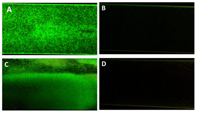
Bacteria and fungi often form biofilms, which are complex communities of microbes bound by an extracellular matrix. Biofilms are beneficial to microbes for various reasons, including adhesion, nutrient cycling, exchange of genetic material, and division of metabolic labor amongst different subpopulations. However, the development of biofilms is often detrimental to humans, such as in the case of plaque accumulation on teeth and medical device infection. Although biofilms can be investigated in traditional cultures, the static nature of the culture can lead to nutrient depletion and metabolite accumulation, which does not reflect natural systems where biofilms modulate their response to environmental changes. Therefore, efficient development of antibiotic and antimicrobial drugs should be done under shear flow conditions.
Figure 4. Confocal Laser Scanning Microscopy of viable P338-adsorbed and unadsorbed biofilms
Conclusions
Accurate investigation of processes that are impacted by flow in the body requires similar flow in vitro to examine relevant responses. Instruments such as BioFlux can create shear stress at specific intensities that can be adjusted to mimic the physiological or pathological shear stress that cells of interest experience in vivo. A key component of the BioFlux system is the ability to provide real-time imaging, enabling a clearer understanding of the interactions between cells or the effects of compounds during drug discovery and development investigations. Adding shear stress to cells in the form of flow can provide many benefits to in vitro experiments that induce physiological responses similar to those that occur in vivo.
References

Lexi Land contributes to developing scientific workflows that are compatible with the CellRaft® AIR System. Her focus at Cell Microsystems involves stem cell research, 3D organoidA 3D, self-organizing cell structure, derived from pluripotent or adult stem cells that mimic aspects of organ function and architecture. work, as well as adherent and suspension cell culture. Lexi received a Bachelor of Science in Biological Sciences from North Carolina State University with a focus in molecular, cellular, and developmental biology.