The satisfaction that comes from seeing a carefully designed experiment unfold and coalesce into a clue, an answer, or a new tool is what makes research and development so gratifying. But every scientist knows that this process is not always seamless. When the goal is big, like Kite Pharma’s ultimate goal of curing cancer, there are bound to be obstacles.
Kite Pharma, a Gilead Company, develops genetically engineered, autologous CAR T cell therapies for the treatment of cancer. Kite’s mission statement emphasizes how these therapies can be used:
“At Kite, our singular focus is cell therapy to treat and potentially cure cancer. Our goal is to bring the promise of cell therapy to as many eligible patients as possible who may benefit. We believe cell therapy has the ability to change the way cancer is treated. Our industry-leading cell therapy technology uses the power of a patient’s own immune system (their white blood cells) to target and attack their cancer cells.”
At the heart of this mission is the ability to engineer cell lines for target discovery and disease modeling. At Kite, the Flow Cytometry and Cell Engineering Core uses Clustered Regularly Interspaced Short Palindromic Repeats (CRISPR) gene editing to develop cell lines for research. Even with expertise and advanced instrumentation at their disposal, the team at Kite found that relying on single cell cloning for cell engineering was causing a bottleneck that was a barrier to efficiency and progress.
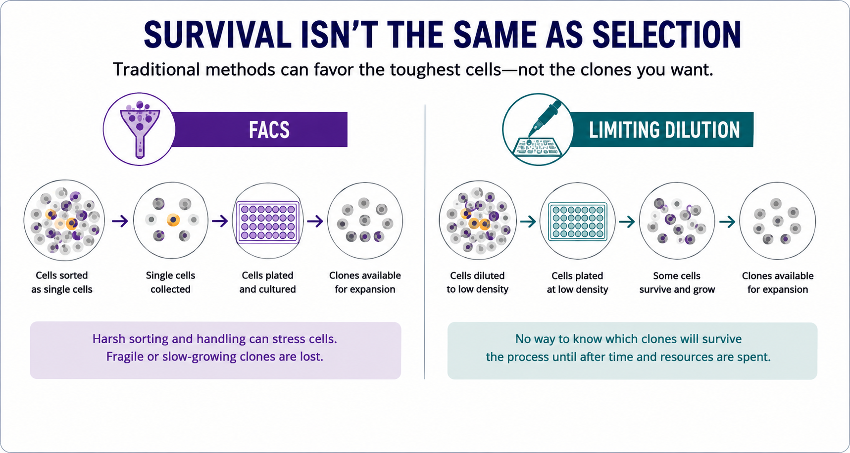
 The Core at Kite Pharma had been using various techniques for single cell cloning, including manual limiting dilutionA statistical cloning approach where cells are diluted to low density to “hopefully” seed one cell per well; commonly used but can be low-efficiency for difficult-to-clone lines., high-speed cell sorting, or combinations of bulk sorting and single cell dispensing. They often encountered poor or variable cell outgrowth after isolation of single cells into 96-well plates, resulting in timelines that were not amenable to their fast-paced goals. To alleviate these pain points and accelerate their cell line development pipeline, they leveraged the CellRaftA microscale polystyrene growth surface within an array used to spatially segregate cells/colonies while maintaining shared media access, enabling imaging over time and targeted isolation. AIR® System from Cell Microsystems.
To compare the efficiency of several methods for cell line generation, Kite created CRISPR knockout Raji cells, which are lymphoblast-like, suspension cells derived from B-lymphocytes commonly used to test anti-tumor activity for therapeutic approaches related to B cell malignancies. Kite analyzed various single cell clone generation workflows using the CellRaft AIR SystemAn all-in-one platform for imaging, identifying, and isolating viable single cells, colonies, or organoids using CellRaft Arrays and software-guided selection with CellRaft Cytometry. as well as traditional cell sorters like the BD FACS Aria Fusion, the SONY SH800 Sorter, and ThermoFisher BigFoot Spectral Cell Sorter.
Concisely stated, cells were transfected with CD19 sgRNAs (Fig. 1), then either sorted directly into 96-well plates using the traditional cell sorters, or bulk sorted to enrich for the CD19PE+ cells and applied to a CellRaft® Array (Fig. 2).


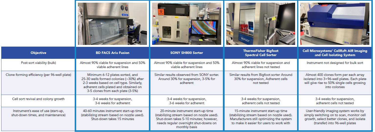
Using the CellRaft AIR to image the CellRaft ArrayConsumables containing thousands of microwells (containing CellRafts) for spatial segregation, imaging, and isolation of single cells, monoclonal colonies, or 3D cultures. over the course of culture, the system captured an image-based record of single cells as they divide into clonal colonies of cells (Fig. 3A-H). The images shown in Fig. 3A-H are representative of individual CellRafts, with Fig. 3A-D and Fig. 3E-H showing the same CellRaft over the course of 4 and 5 days, respectively. After isolation, Kite found that colonies isolated from the CellRaft Array grew out in over 60% of wells, compared to only 30% growth from single cells dispensed into 96-well plates using cell sorters.

Ultimately, the team at Kite Pharma implemented CellRaft Technology in their Core because it provides a greater number of clones due to superior cell growth and expansion, enabling them to shorten their timeline to cell line generation and improve their efficiency. Combined with the ease-of-use, confirmation of monoclonalityEvidence that a colony/line originated from one cell; often supported by time-course imaging and traceability., and software-driven clone selection, CellRaft Technology enables a more researcher-friendly workflow that helps companies achieve their overarching goals.



Dr. Megan Harrison is responsible for demonstrating the power of the CellRaft® AIR System to the scientific community, training new users on CellRaft® Technology, and supporting current users by helping them to develop new workflows and to optimize existing processes. Dr. Harrison has experience as a researcher and leader in industry and academic settings. Most recently, as Principal Scientist for Frequency Therapeutics, she developed and characterized 3D culture models, such as primary tissue-derived organoids and spheroids, which were used for small molecule drug discovery in the regenerative medicine space. Dr. Harrison received post-doctoral training at Yale School of Medicine in the Cell Biology department, where her research focused on endosomal trafficking. She earned her Ph.D. in Pathobiology from The Pennsylvania State University, where her research focused on Paramyxovirus budding. Dr. Harrison holds a B.S. in Pre-Veterinary Medicine and Animal Biotechnology from the University of Delaware, where she first learned to conduct research in avian virology.