
Adhesion Strength
The adhesion of planktonic microbes and their subsequent development into biofilms is dependent upon several factors, including shear stressThe tangential force per unit area from flowing fluid; a key parameter in vascular biology, platelet function, leukocyte rolling, and biofilm adhesion studies., nutrient availability, and the composition of the attachment surface. Examination of the microbial adhesion and growth processes enables the investigation of antibiofilm and antimicrobial surface treatments.
BioFlux shear flow systems enable high-throughput visualization and analysis of all the steps of microbial adhesion and growth. Controlled shear flowFluid movement that applies force to cells and biofilms; used to mimic physiological/pathological conditions. can be used to treat biofilms with antimicrobial compounds, biosurfactants, or other solutions that alter adhesion. Furthermore, the microfluidic channels of BioFlux plates can be coated with an antibiofilm treatment of interest or custom substrate plates can be leveraged for microbiology investigations.
BioFlux microbial adhesion workflow
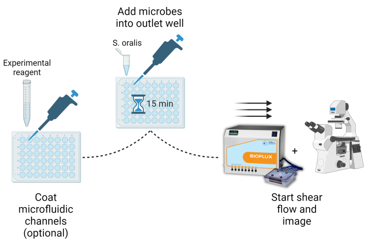
Biofilm adhesion using BioFlux
Role of Force-Sensitive Amyloid-Like Interactions in Fungal Catch Bonding and Biofilms
Real-time adhesion of Als5p-expressing Candida albicans cells under a shear flow rate of 0.8 dyn/cm².
High-Throughput Microfluidic Method To Study Biofilm Formation and Host-Pathogen Interactions in Pathogenic Escherichia coli
Time-lapse growth of Escherichia coli (strain EHEC EDL933) on HRT-18 monolayers in a BioFlux plate.
Publication spotlight: Role of Force-Sensitive Amyloid-Like Interactions in Fungal Catch Bonding and Biofilms
Key BioFlux Advantages For Adhesion Strength Studies
- Accurately control shear forces to quantify bacterial adhesion
- Microfluidic design allows for minimal media usage, contributing to longer experiments and more time for shear-enhanced flow analysis
- Use biosurfactants, antimicrobial compounds, etc. to inhibit biofilm formation and adherence under flow
- Perform binding assays and measure the binding specificity of microbial surface proteins in a microfluidic channel
Count Microbes With Confidence
CASY Cell Counter & Analyzer

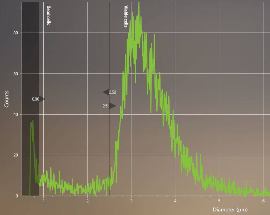
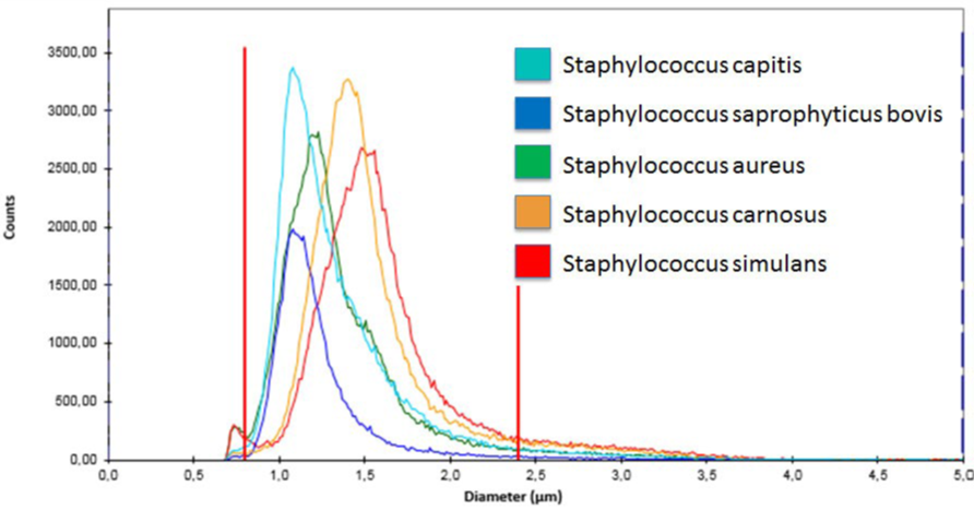
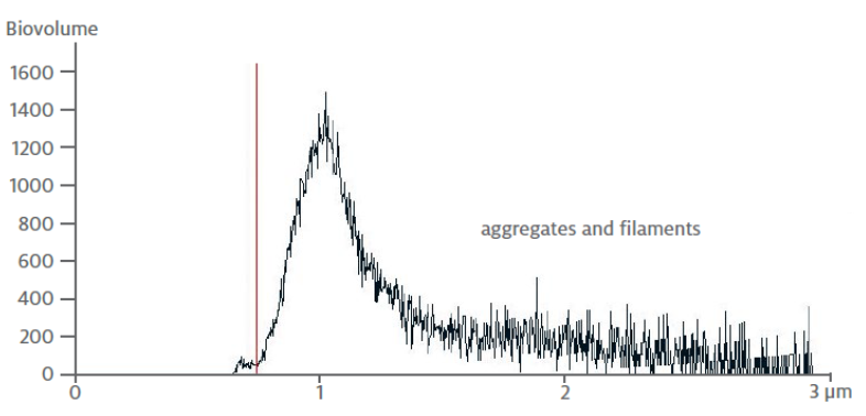
Benefits of CASY for Microbiologists
-
Accurate mixed-species counting
Count up to 5 species of microbes from heterogeneous samples, all at once -
Count almost anything
A 0.7-120 µm measurement range provides unmatched counting flexibility -
Assess aggregation
Volume-based aggregation correction provides accurate aggregate counting and changes in the level of aggregation -
Spore differentiation
Accurately discriminate between haploid and diploid spores -
Measure cells with flagella
1 MHz detection enables precise volume-based size determination of elongated cells -
Biomass, proliferation, & biovolume all at once
Measure cell volume and counts simultaneously to monitor growth
FAQs:
Microbial Adhesion Using the BioFlux
What factors influence microbial adhesion and biofilm formation?
Microbial adhesion depends on shear stress, nutrient availability, and surface chemistry. Under dynamic shear flow, planktonic microbes initially attach, then develop into biofilms. Understanding these stages is essential for evaluating antimicrobial surfaces or adhesion inhibitors. The BioFlux microfluidic platform enables real-time analysis of these processes under controllable shear stress.
How does shear flow impact measurement of bacterial adhesion strength?
Shear flow tests the force required to detach adherent microbes. The BioFlux applies precisely controlled laminar shear across microfluidic channels embedded in BioFlux plates, allowing quantitative measurement of adhesion strength and a direct comparison of surface treatments or compounds.
Can the BioFlux enhance throughput for microbial adhesion testing?
The BioFlux employs well-plate integrated microfluidic channels that enable the delivery of precise, programmable shear flow while simultaneously testing different shear stress rates and coatings—ideal for rapid screening of antibiofilm or anti-adhesion agents.
Can the BioFlux simulate physiological conditions for microbial adhesion studies?
Yes. The BioFlux replicates in vivo shear forces across a 0–200 dyn/cm² range, simulating physiological environments such as blood flow, saliva, or urinary tract conditions.
How does the BioFlux aid in evaluating anti-adhesion surface coatings?
Microfluidic channels can be pre-coated with custom materials or antibiofilm agents. Under shear, microbial attachment and detachment can be visualized, quantifying adhesion strength variations.
Which pathogens have been studied for adhesion using BioFlux?
The BioFlux has been used to examine the adhesion of fungal and bacterial species, such as Candida albicans and Escherichia coli among others, on epithelial cell monolayers.
How does BioFlux compare with traditional batch adhesion assays?
Unlike static assays in microplates, the BioFlux offers image-based, real-time analysis of biofilmA structured microbial community attached to a surface, typically more tolerant to antibiotics/biocides than planktonic cells. adhesion, propagation and detachment under controlled shear flow. This provides quantitative adhesion strength data and mimics physiological conditions, making it more predictive and reproducible.
Is the BioFlux compatible with live-cell imaging for adhesion studies?
Yes. BioFlux plates feature glass bottom microfluidic channels compatible with inverted microscopes. Researchers can monitor real-time adhesion, detachment, and biofilm evolution with high-resolution imaging under physiological flow.