
White Paper
Development of Monoclonal Cell Lines - Available Technologies and Overcoming Challenges

Introduction
Single-cell cloning is the process by which a cell line is generated from a single starting cell that has been segregated from a heterogeneous population, typically one that has been prepared from transfections, transductions, or primary cells from tissue or biopsy.1 As new methodologies evolve, life science drug development has begun to expand from small molecules toward more specific biologics and advanced therapy medicinal products (ATMPs). Single-cell workflows have played an important role in this expansion, as they have clinical and scientific impacts on crucial cross-disciplinary applications in multi-omics,2 immune oncology,3 rare cell identification,4 drug discovery,5 stem cells (regenerative medicine),4,6,7 biologics,8 organoids,9,10 and epigenetics.11,12
In recent years, cell line development for the purpose of analytics, bioproduction and therapeutics has evolved into a crucial workflow, with monoclonalityEvidence that a colony/line originated from one cell; often supported by time-course imaging and traceability. as a critical requirement (Figure 1). The development of more specific therapies for a diverse range of disease conditions has also driven the advancement in single-cell workflow particularly cloning which has become a vital requirement in biopharmaceutical research and product development.
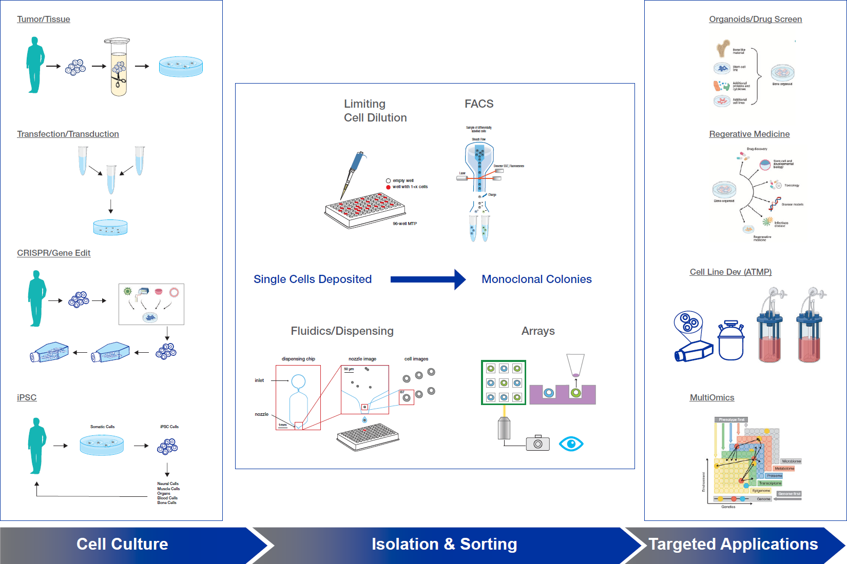
Current Methods
Currently, the two widely used single-cell isolation techniques are limiting dilutionA statistical cloning approach where cells are diluted to low density to “hopefully” seed one cell per well; commonly used but can be low-efficiency for difficult-to-clone lines. and single cell dispensing, sometimes aided by fluorescence cell sorting. Although perceived to be commonplace, these techniques are tedious and time-consuming, and have other significant drawbacks including requiring stringent sample preparation and extensive training and equipment maintenance, and typically necessitating serial rounds of cloning to produce a single monoclonal cell line.13 These disadvantages also often result in poor cell viability and proliferation.
Despite the drawbacks, the most convenient and cost-effective single-cell isolation technique is limiting dilution.14,15 This involves the generation of a monoclonal line from a polyclonal pool through manual serial dilutions, image analysis of individual clones, and subsequent expansion of clones of interest. While the ability to obtain individual cells from diluted cell suspensions seems simple and straightforward when employing hand pipettes or automated robotic pipetting platforms, it is often difficult to retrieve a sufficient number of the desired monoclones. At the end of this process, it is not unusual to have wells with multiple cells, or no cells at all, and a lack of viable clones is often an additional problem. Moreover, given that it is difficult to determine if cells were truly isolated before expansion, there is no guarantee that the subsequent colonies originate from single cells.16
The use of flow cytometry and cell sorting is another technique that leads to a false impression of improvement upon limiting dilution.14,17 Here, cells are analyzed and sorted based on hydrodynamic focusing phenomenon and specific cellular characteristics. Fluorescence-activated cell sorting (FACS) enables a single cell to be separated from a cell suspension with some degree of purity. In fact, the cell sorter is capable of placing a single cell in a microplate well with reasonable accuracy and efficiency. Benchtop cell sorting systems with multiple lasers and colors can improve the precision and speed of the sorting and selection process.18 However, this necessitates significant gating requirements and setup for every population to be sorted, to ensure retrieval of the maximal number of viable clones.19 In some cases, limited by the availability of fluorescently tagged antibodies developed towards surface epitopes, the preparation of reagents can become a highly complicated exercise. Furthermore, the cell sorting and dispensing process can damage the cells, altering their metabolic state and inducing oxidative stress.20 In fact, due to the fraction of FACS-sorted cells that are dead or have impeded growth, it is common for only 20% of isolated single cells to produce usable colonies.19,21 Cells subjected to FACS-sorting show a 50% increase in reactive oxygen species, suggesting that as these cells transition from proliferating in cell culture to the stressful environment of sorting and dispensing, their metabolism shifts from a state of anabolism to catabolism. Such a metabolic transition decreases reductive biosynthetic reactions utilizing NADPH, and cells undergoing this transition switch on apoptotic genes or shutdown S phase synthesis.20 As a result, diminished outgrowth is often reported in single cell progenitors initiated via FACS, and necessary outgrowth yields tend to take longer to achieve.
Although these techniques are commonly used across academic and the biopharmaceutical industries, there are major drawbacks. First, the cell preparation must be a single-cell suspensionA uniform mixture of individual cells, required for accurate counting, many microfluidic assays, and single-cell sequencing. and cannot be used for adherent cells without extensive enzymatic or physical treatment. Second, both methods rely on statistical probabilities to claim monoclonality, require multiple serial rounds of cloning to produce a clonal cell line, and often overestimate the probability of monoclonality due to the presence of cell clusters21. Thus, the verification of a single cell progenitor is a tedious, time-consuming, microscopic examination of all microplate wells and does not provide a clearly documented image of a single cell. Therefore, proving monoclonality is laborious and difficult to demonstrate with these techniques. Systems for these methods of single-cell clone generation currently consist of multiple platforms, components, and steps. This can be economically taxing, limits compatibility and availability, and often requires troubleshooting at different stages of the workflow, adding another layer of complexity to this already demanding process (Table 1).
Table 1: Cell Sorting and Isolation Methods
| Isolation Methods | Description | Advantages | Disadvantages | Cost |
|---|---|---|---|---|
| Limiting Dilution – Manual | Serial dilution until solution is statistically calculated to be one cell per microliter | Established, simple and familiar protocols; perceived to be low cost | High failure rate; error prone; tedious; extended experimental timeline (~10 weeks); additional equipment/space required (biosafety cabinet, incubator, cell counter); contamination risk; high risk of isolating multiple cells; manipulation of cells can perturb expression profiles; not designed for bulk sorting | $ |
| Limiting Dilution – Automated | Robotic-controlled micropipettes | High accuracy; fluorescence can be used | Lack of software analysis increase time and effort to get reasonable results for isolation & selection | $$ |
| Flow Sorting | Microdroplets with single cells are isolated by electric charge at high pressure | Enables bulk or single-cell sorting high accuracy and precision for identifying cells/populations of interest; fluorescent markers can be used to isolate sub-populations. | Requires separate single-cell dispenser; low yield; requires off-platform propagation for cell line development; not amenable to organoid/3D biology; equipment and manual labor- requires hands on attention; fl uidics perturb cell metabolism; perturb expression profiles and damage cells | $$ |
| Microfl uidic platforms | Microfluidic chips isolate single cells in flow channels | High-throughput; reactions can be performed on-chip; reduced reagent costs | High failure rate; prone to contamination; highly complicated fl uid mechanics can complicate outcomes; lack of imaging options | $$ |
| Cell Dispensing (droplet) | Single cell trapped in microfl uidic drops | Single cells can be imaged in a fl ow path | Highly complicated fl uid mechanics can complicate outcomes | $$ |
| Cell Raft Technology | Single cells grown in specialized culture dish; one single platform for integrated imaging, analysis, isolation. | Able to isolate up to 400 clones from each array into 96-well plates, with each plate giving rise to >90% single-cell growth into colonies; can propagate stem cells/iPSCs, organoids, or screen T-cells; fast ease of tracking and tracing of clonal propagation unique powerful software drives selection and isolation of cells based on highly specific end user requirements | Not ideal for high-throughput single cell genomics; not designed for bulk sorting; cannot be integrated with sample prep methodologies | $ |
| Optofl uidic Technology | Uses light and millions of light-actuated pixels to move individual cells so they can be isolated, cultured, assayed, and exported | Integrated workfl ow | Limited number of cells; chip only has 5,000 positions; not every nanopen position is occupied; very limited applications; requires a fully dedicated lab technician to operate; technology has not been fully adopted yet | $$$$$$ |
Recently, several new products specializing in cell sorting and dispensing have emerged, each marketing their system as a method for streamlining the generation of single-cell clones. These setups vary widely in specifications and cost, ranging from $150,000 to millions of dollars.
The most common of these systems are cell dispensers. These platforms use microfluidics combined with brightfield imaging or fluorescence detection to deposit a single cell in one well of a 96- or 384-well collection plate. These platforms seek to improve upon limiting dilution techniques by eliminating the time and resources spent serially processing culture plates by directly obtaining an isolated single cell. This may provide some improvements over limiting dilution and improved confidence in monoclonality; however, such systems have limited benefits. Outgrowth of the isolated single cells must be performed separately, necessitating additional equipment, resources, and space demands. This leads to additional costs beyond the platform itself, which alone can run from several hundreds of thousands to millions of dollars in purchase price. The process of physical manipulation in fluidic channels and droplet impact of dispensing can also harm the isolated cell, perturbing expression profiles and reducing outgrowth (Figure 2).22 Furthermore, these platforms are specific in function and do not support other forms of selection or propagation, and therefore those seeking single-cell workflows to propagate stem cells/iPSCs, organoids, or screen T-cells cannot make use of them. Thus, although single-cell dispensers represent an improvement over traditional manual methods, they do not meet all needs to facilitate rapid and efficient development of clones.
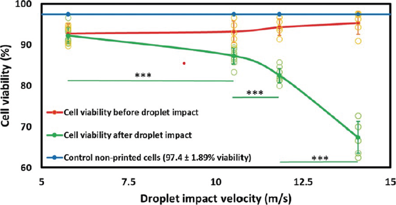
(adapted from Ng et al., 2022)
With the market saturated with a variety of platforms, the researchers are left to navigate and decide which system is most appropriate and cost-effective for their needs. While new techniques such as microfluidic platforms and automated clone pickers show promise for the screening, selection, and isolation of monoclones, they still fall short on delivering on a high volume of viable monoclonal colonies without constraining resources. There is a clear need for more comprehensive tools that encompass the entire workflow from single-cell separation through outgrowth, and that are applicable for multiple cell lines and types.
Overcoming the Challenges
Based on the current needs of researchers (Table 2) and methods described, it is clear that a system or technology is needed to obtain single cells while providing natural conditions to maintain cell cycle kinetics and ensure that bio-chemical equilibrium is not perturbed during the process of isolation. New technologies have been developed to meet some of these unmet needs.
Table 2: Comparison of the different technologies from the perspective of an experimental workflow.
| Experimental Needs | Limited Dilution (manual) | Limited Dilution (automated) | Flow Sorting | Cell Dispensing (droplet) | Optofluidic Technology | CellRaft Technology |
|---|---|---|---|---|---|---|
| Main Applications | Single Cell Cloning | Single Cell Cloning | Single Cell Isolation | Single Cell-Omics; Cell Line Development; Gene Therapy | Clonal Cell Line Development | Cell Line Development Single Cell cloning; iPSC & Organoids Engineering, Development and Maturation |
| Throughput (number of cells for single cell propagation) | Low | High | High | High | High | Medium |
| Cell Viability | Very Low | Very Low | Low | Low | High | Very High |
| Outgrowth Efficiency | Negligible | Very Low | Medium | Medium | Low | Very High |
| Visual Control | None | None | None | Partial | Yes | Yes |
| Cell Selection | None | None | Limited | Limited | Limited | Yes fully capable |
| Starting Number of Cells Needed | High | High | Moderate | Moderate | Low | Moderate |
| Flexibility (Own Protocols) | Yes | Yes | No | No | No | Yes |
| Software | None | Used for Robotic Control | Analyze, Segregate | Analyze, Segregate | Analyze, Robotic Control | Analyze, Isolation, Monitoring as a Function of Time |
| Lab Skills Needed | Low | Low | High | Low | Very High | Low |
| Integration with Lab Management System | No | Yes | Yes | No | Yes | No |
| Integrated Workflow | No | No | No | No | Yes | Yes |
| Footprint in the Lab | Negligible | Large | Large | Small | Very Large | Small |
| Number of Cell Types Demonstrated | NA | ~10-25 | >100 | <20 | 2 to 1 | ~100 |
| 2D or 3D Biology | No | No | No | No | No | Yes |
| Robotic Compatible | None | Yes | Some | No | Yes | No |
| Real Time Live Cell Image Analysis | No | No | No | Yes | No | No |
| Track and Trace (Time Course/Audit Trail) | No | No | Limited | Limited | Yes | Yes |
| Image versus Signal | None | Image | Signal | Image | Image | Image |
| Proof of Monoclonality | Indirect | Indirect | Indirect | Indirect | Direct | Direct |
The CellRaft® Technology (Figure 3) provides flask-like culture conditions at the resolution of a single cell, with gentle and automated isolation using image-based attributes for function, gene expression, and morphology. This technology is manifested in the CellRaftA microscale polystyrene growth surface within an array used to spatially segregate cells/colonies while maintaining shared media access, enabling imaging over time and targeted isolation. AIR® System23, which is an integrated platform for growing, scanning, analyzing, and isolating single-cell derived monoclonal colonies. This system relies on the CellRaft® Array, which is a cell culture dish with 10,000-150,000 microwells called CellRafts. This design allows the cells to settle gently by gravity and distribute across the array into a variety of single, double, or clustered combinations. According to the Poisson distribution model, approximately 40% – 60% of CellRafts are populated by single cells, depending on seeding density. The unique design allows all the cells to share the same media and extracellular growth factors or cytokines, mimicking the growing conditions of an actual flask or a reactor. This allows physical isolation of single cells without physical perturbation and eliminates any biochemical or physiological changes.

The CellRaft AIR SystemAn all-in-one platform for imaging, identifying, and isolating viable single cells, colonies, or organoids using CellRaft Arrays and software-guided selection with CellRaft Cytometry. enables the ability to individually image and analyze the cells in brightfield or fluorescent imaging modalities. The CellRaft Cytometry™ software allows selection of clones that accurately fit the attributes defined by the end user or application. Acquisition, isolation, and retrieval of monoclonal colonies are performed automatically on the same platform by the mechanical actuation of a magnetic wand, which gently transfers the colony-containing CellRaft to a 96-well collection plate.
The CellRaft AIR System can image, track, analyze, and automate the isolation of colonies from single cells using one instrument with no minimum sample size requirement. Due to the fact that single-cells are grown without microfluidic separation or perturbation ensures viability and vitality of single-cells and allows them to develop into healthy clones. The CellRaft ArrayConsumables containing thousands of microwells (containing CellRafts) for spatial segregation, imaging, and isolation of single cells, monoclonal colonies, or 3D cultures. relies on shared media across the array allowing cell-to-cell communication during clonal development, making it possible to obtain 10X to 50X more viable, highly proliferative monoclonal colonies. This is a significant improvement over other systems, as the flask-like conditions help promote the existence and selection of cells that will not be in a resting phase, early apoptosis, senescent, or other conditions that make the cell difficult or impossible to propagate after isolation.19 Moreover, the CellRaft AIR System is highly versatile in that it can achieve colony growth from primary, adherent, or suspension cells, including iPSCs and immune cells. Additionally, users can grow iPSCs into 3D cell systems, including organoids, for applications such as cancer immunology, multi-omics and cellular heterogeneity studies.
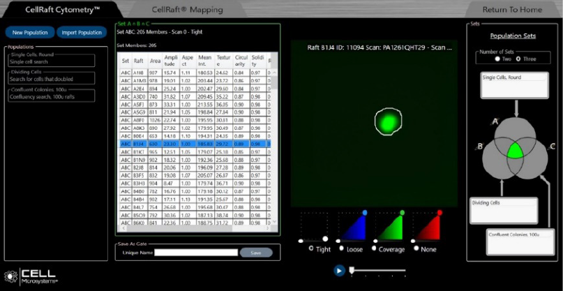
The “brains” of the CellRaft AIR System is the software called CellRaft Cytometry that selects, scans and images thousands of CellRafts, enabling automated identification and isolation of the desired clones (Figure 4). The software allows users to easily interact with thousands of viable cells in real time. It seamlessly integrates with the hardware with intuitive navigation of the many features included. The software can be used on and off the system allowing the user to analyze cellular data on a desktop or laptop. The key features of the CellRaft Cytometry softwareCompanion software to the CellRaft AIR instrument enabling image analysis of cells, including verifying single cells and performing phenotypic selection, for mapping CellRafts for automated isolation. include (1) versatility – multiparameter analysis (time, morphology, phenotype); (2) automated CellRaft identification and isolation, resulting in software guided biology decisions; (3) unbiased CellRaft selection, reducing errors in identification; (4) easy template creation with a QuickStart library; (5) savable user-defined parameters for assay accuracy and consistency; (6) track and trace capability for an audit trail; and (7) simultaneous scanning and data analysis in real time.
Workflow
Similar to standard culturing methods, cells are plated on the CellRaft Array which is then loaded into the CellRaft AIR System platform. Cells are imaged with three-channel fluorescence and brightfield microscopy and sorted with user-defined thresholds, filtering, and gating (Figure 4). The software will scan for expression, time, morphology, and automatically isolate the desired CellRaft based on user criteria.24 The system can isolate a full 96-well plate of individual, undisturbed cells, or colonies in under an hour for expansion and downstream analysis.25
The CellRaft Air System offers a number of advantages over other currently available systems. It provides a more efficient single-cell workflow, including clonal colony propagation, by combining imaging, identifying, and isolating in one instrument and on one consumable. It achieves this while also producing very high clonal yields with robust viability and cell proliferation. This is a significant improvement over other platforms, which often have additional space and instrument needs. Furthermore, automation of all steps provides significant benefits, including real time imaging, identification, and isolation of cells and small colonies for outgrowth in 96-well plates (Figure 3). This not only accelerates the timeline to results, but also reduces costs and contamination risks. The system includes several onboard assays,25 including cell characterization, co-culturing, cell-drug, and cell-cell interactions. Finally, unlike other systems, the CellRaft AIR System supports a wide range cell types and 3D cell systems,25 including stem cells (including iPSCs), animal or human cells, primary cells, immortalized cells, adherent cell, suspension cells, and organoids.
Recent experimental and cost analyses including hands on time to run the protocol between limiting dilution and CellRaft Technology indicates that the latter delivers a high return on investment in terms of outgrowth efficiency, time and cost as seen in Table 3.
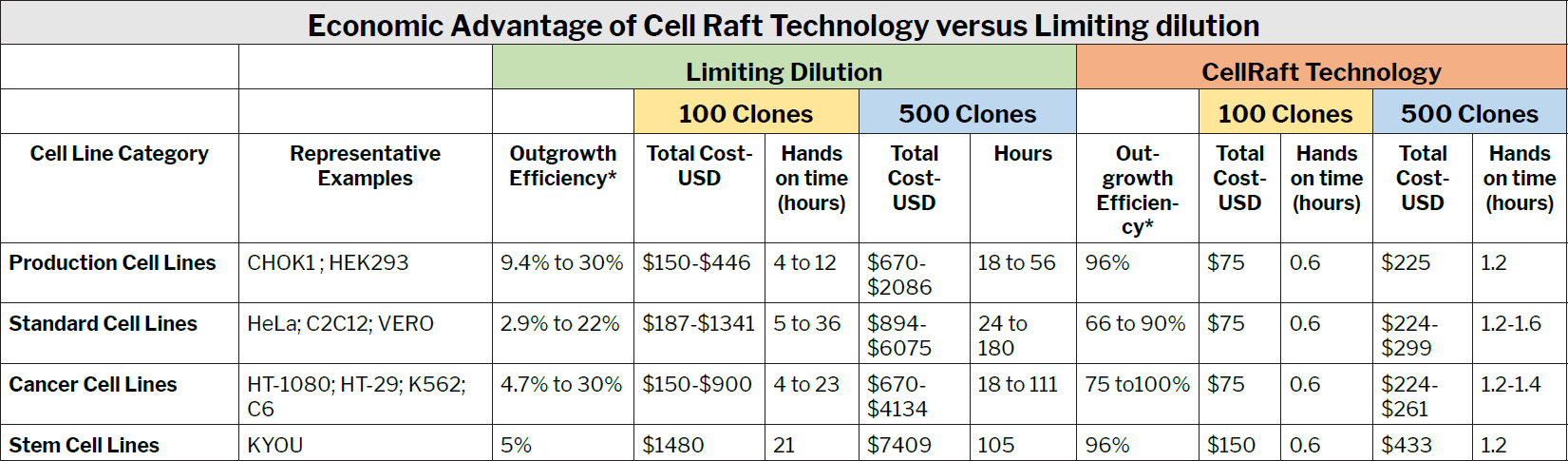
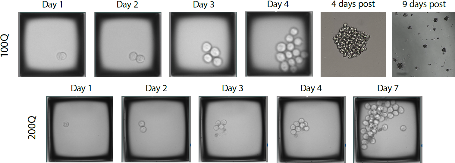
Currently single cell workflows for approximately 100 different cell lines and types have been successfully demonstrated on the CellRaft AIR System (Figure 5), and new workflows are being constantly developed and validated.
Conclusions
References
- Kline, C. The art of generating single cell clones. BiteSize Bio
https://bitesizebio.com/42870/to-be-a-clone-or-multiclonal/ (2019). - Mincarelli, L., Lister, A., Lipscombe, J. & Macaulay, I. C. Defining Cell Identity with Single-Cell Omics. Proteomics 18, 1700312 (2018).
- Gohil, S. H., Iorgulescu, J. B., Braun, D. A., Keskin, D. B. & Livak, K. J. Applying high-dimensional single-cell technologies to the analysis of cancer immunotherapy. Nature Reviews Clinical Oncology 2020 18:4 18, 244–256 (2020).
- Gach, P. C., Attayek, P. J., Herrera, G., Yeh, J. J. & Allbritton, N. L. Isolation and in vitro culture of rare cancer stem cells from patient-derived xenografts of pancreatic ductal adenocarcinoma. Anal Chem 85, 7271–7278 (2013).
- Heath, J. R., Ribas, A. & Mischel, P. S. Single-cell analysis tools for drug discovery and development. Nature Reviews Drug Discovery 2015 15:3 15, 204–216 (2015).
- Zheng, H. et al. Single-cell analysis reveals cancer stem cell heterogeneity in hepatocellular carcinoma. Hepatology 68, 127–140 (2018).
- Kester, L. & van Oudenaarden, A. Single-Cell Transcriptomics Meets Lineage Tracing. Cell Stem Cell 23, 166–179 (2018).
- Diep, J. et al. Microfluidic chip-based single-cell cloning to accelerate biologic production timelines. Biotechnol Prog 37, e3192 (2021).
- Brazovskaja, A., Treutlein, B. & Camp, J. G. High-throughput single-cell transcriptomics on organoids. Curr Opin Biotechnol 55, 167–171 (2019).
- Youk, J., Kwon, H. W., Kim, R. & Ju, Y. S. Dissecting single-cell genomes through the clonal organoidAn organoid derived from a single cell. technique. Experimental & Molecular Medicine 2021 53:10 53, 1503–1511 (2021).
- Tedesco, M. et al. Chromatin Velocity reveals epigenetic dynamics by single-cell profiling of heterochromatin and euchromatin. Nature Biotechnology 2021 40:2 40, 235–244 (2021).
- Meir, Z., Mukamel, Z., Chomsky, E., Lifshitz, A. & Tanay, A. Single-cell analysis of clonal maintenance of transcriptional and epigenetic states in cancer cells. Nature Genetics 2020 52:7 52, 709–718 (2020).
- Riba, J., Schoendube, J., Zimmermann, S., Koltay, P. & Zengerle, R. Single-cell dispensing and ‘real-time’ cell classification using convolutional neural networks for higher efficiency in single-cell cloning. Scientific Reports 2020 10:1 10, 1–9 (2020).
- Ye, M., Wilhelm, M., Gentschev, I. & Szalay, A. A Modified Limiting Dilution Method for Monoclonal Stable Cell Line Selection Using a Real-Time Fluorescence Imaging System: A Practical Workflow and Advanced Applications. Methods Protoc 4, 1–14 (2021).
- Lefkovits, I. & Waldmann, H. Limiting dilution analysis of the cells of immune system I. The clonal basis of the immune response. Immunol Today 5, 265–268 (1984).
- Evans, K. et al. Assurance of monoclonality in one round of cloning through cell sorting for single cell deposition coupled with high resolution cell imaging. Biotechnol Prog 31, 1172 (2015).
- Welch, J. T. & Arden, N. S. Considering “clonality”: A regulatory perspective on the importance of the clonal derivation of mammalian cell banks in biopharmaceutical development. Biologicals 62, 16–21 (2019).
- Benchtop Microfluidic Cell Sorter: WOLF G2 | NanoCellect.
https://nanocellect.com/products/wolf-g2-cell-sorter/?utm_campaign=GS_Brand_BMM&utm_source=google&utm_medium=cpc&utm_content=text&utm_term=wolf%20nanocellect&utm_campaign=GS_Brand_BMM&utm_source=adwords&utm_medium=ppc&hsa_acc=1686850884&hsa_net=adwords&hsa_grp=77679615039&hsa_ver=3&hsa_kw=wolf%20nanocellect&hsa_tgt=kwd-819603803256&hsa_mt=e&hsa_ad=385078632457&hsa_src=g&hsa_cam=6542195443&gclid=Cj0KCQjwio6XBhCMARIsAC0u9aE1LpS4E3bgGVwZ6b422n1-eCCdJ7TfRhMciUHFhpuAIxWG6rW1wLEaApHvEALw_wcB. - Riba, J., Schoendube, J., Zimmermann, S., Koltay, P. & Zengerle, R. Single-cell dispensing and ‘real-time’ cell classification using convolutional neural networks for higher efficiency in single-cell cloning. Scientific Reports 2020 10:1 10, 1–9 (2020).
- Llufrio, E. M., Wang, L., Naser, F. J. & Patti, G. J. Sorting cells alters their redox state and cellular metabolome. Redox Biol 16, 381–387 (2018).
- Evans, K. et al. Assurance of monoclonality in one round of cloning through cell sorting for single cell deposition coupled with high resolution cell imaging. Biotechnol Prog 31, 1172–1178 (2015).
- Ng, W. L. et al. Controlling Droplet Impact Velocity and Droplet Volume: Key Factors to Achieving High Cell Viability in Sub-Nanoliter Droplet-based Bioprinting. Int J Bioprint 8, 1–17 (2022).
- The CellRaft AIR System enables workflows to image, sort and isolate viable single cells.
https://cellmicrosystems.com/cellraft-air-systems/. - Cell Microsystems – CytoSort Arrays.
https://cellmicrosystems.com/cellraft-products/cytosort-arrays/. - Cell Microsystem. CellRaft AIR System Brochure. (2021).