
The advancement of 3D culture systems has transformed cell-based assays for biological research and drug discovery due to their ability to re-capitulate the structure and cellular complexity of in vivo tissues. Organoids are unique due in their ability to self-organize and closely replicate in vivo pathophysiology. Most laboratory instruments employed to assess endpoints using traditional organoid cultures have limitations that preclude assessment of the heterogeneity within the population or retrieval of single, intact 3D structures for downstream applications.

The AIR System offers unique advantages over technologies, such as flow cytometry and droplet dispensers, designed to assess cells in suspension. The limitations to other technologies include impacts on cell viability, inability to interrogate small numbers of cells, and reliance on fluorescent markers or staining for cell characterization. To demonstrate the value of the CellRaft technology for use in suspension cell line development, we have established methodologies for attaching suspension cells to the CellRaft Arrays during single cell expansion while still allowing the cells to expand in suspension after the clone of interest has been isolated.
Therapeutic proteins play an essential role in the biological pharmaceutical market and are used in the treatment of many diseases, such as diabetes, cancer, and anemia. One of the main goals in recombinant protein development is the establishment of high-quality monoclonal cell lines that consistently express large amounts of the given protein. Chinese hamster ovary (CHO) cell lines have dominated the industry as commercial hosts for recombinant protein production; however, the process of generating a homogenous CHO cell line is not trivial.

Gene editing workflows require transformation of a large number of cells, followed by isolation of individual cells from the larger population to establish clonal colonies. Given the large number of gene edits required for contemporary research and the labor-intensive components of the workflow, there is an unmet need to automate post-transformation cloning.

Functional genomic analysis provides a connection between cellular phenotypes, such as drug responsiveness, and their underlying genomic precursors, such as sequence variation or differential gene expression.
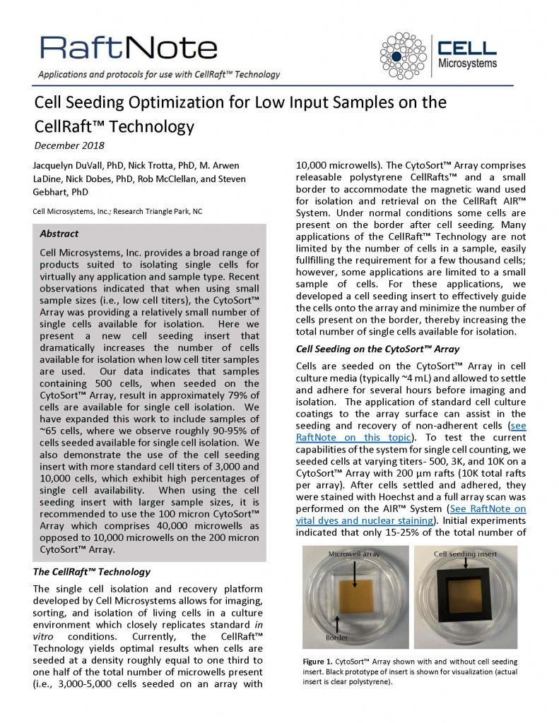
One of the benefits of the CellRaft Technology is the isolation and recovery of single cells even from low cell titer samples. This RaftNote describes a new cell seeding insert that significantly increases the number of cells available for isolation when low cell titer samples are used.

Sorting and isolating single cells is a key sample preparation step in many contemporary workflows including genome editing by CRISPR/Cas9, single cell genomics and differentiation of induced pluripotent stem cells (IPSCs) along with a broad range of other methods. Here, we present optimized methods for staining live cells on the Cell Microsystems CellRaft Array, a microwell array with releasable single cell isolation features called CellRafts.
