As drug discovery and disease modeling become increasingly sophisticated, 3D cell culture has emerged as a powerful alternative to traditional 2D assays and animal models. In a recent webinar Dr. Allysa Stern showcased how researchers are using the CERO 3D Incubator and Bioreactor to streamline development of organoidA 3D, self-organizing cell structure, derived from pluripotent or adult stem cells that mimic aspects of organ function and architecture. models that are scalable, reproducible, and biologically relevant.
Why 3D Matters Now More Than Ever
Despite decades of optimization in drug development, the failure rate for candidate therapeutics remains high, often due to poor translation from animal models to human biology. That’s why researchers are shifting toward more predictive, human-relevant systems like organoids derived from induced pluripotent stem cells (iPSCs). These models better capture tissue-specific physiology and patient diversity, offering an edge in everything from disease modeling to toxicity screening.
Introducing the CERO 3D
The CERO 3D is a benchtop bioreactor system that enables gentle, dynamic suspension culture of spheroids and organoids. Designed for ease of use, it supports up to four independently controlled tubes, allowing researchers to optimize protocols in parallel. Unlike traditional bioreactors, the CERO 3D requires no impellers or external fluidics. Instead, it uses integrated baffles to create a low-shear, homogenized environment ideal for 3D culture.
Case Studies in 3D Organoid Development
1. Cerebral Organoids for Brain Cancer Modeling
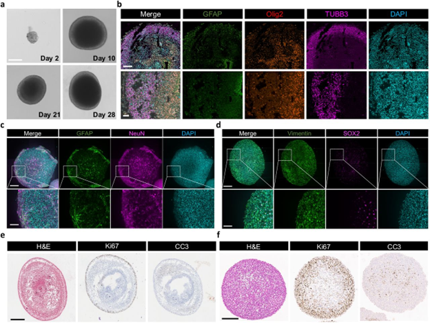
Researchers used the CERO 3D to culture neurospheres and cerebral organoids from adult neural stem cells (Wolfram et al., 2025). Compared to orbital shaker methods, the CERO 3D yielded organoids with better morphology, longer viability (up to 5 months), and greater structural fidelity (Figure 1). These organoids were also used to model glioblastoma, achieving a 5x increase in assembloid surface connectivity, an important factor in accurately representing the tumor microenvironment.
Figure 1. aNSC-derived large cerebral organoid culture. a, Growth stages of cerebral organoids derived from aNSCs at 2, 14, 21 and 28 days. Scale bar: 200 μm. b, Thin sections of cerebral organoids showing neural maturation markers for astrocytes (GFAP), neurons (TUBB3) and oligodendrocyte lineage cells (Olig2). Scale bar: 200 μm, 50 μm. c and d, IF staining of whole 8-week-old organoids showing differentiation markers GFAP and NeuN for astrocytes and neurons (c) and neural stem cell markers Vimentin and SOX2 (d). Scale bars: 225 μm, 100 μm. e and f, IHC analysis of 12-week-old organoid sections, stained for Ki67 and CC3 in orbital shaker (e) and CERO (f) cultures. Scale bars: 500 μm (e) and 300 μm (f).
2. Cardiac Organoids for Cardiotoxicity Screening
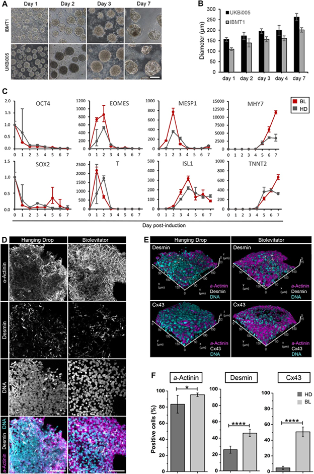
Using two iPSC lines, researchers differentiated cardiomyocytes in the CERO 3D and found faster mesoderm and cardiac lineage specification compared to hanging drop culture (Fischer et al., 2018; Figure 2C). The resulting cardiospheres expressed higher levels of structural and functional markers, including a >10x increase in connexin-43, which supports synchronized contraction (Figure 2E,F). Remarkably, one CERO tube outperformed 10+ flasks or 100 96-well plates in total cardiomyocyte yield.
Figure 2. 3D cardiac induction of hiPSCs in static or dynamic conditions. Cardiospheres differentiated in the BL (CERO 3D) from two hiPSC lines (UKBi005 and IBMT1) were harvested on different days (A) and their diameter was measured (B; Mean ± SEM; n = 20/day). The expression of pluripotency (OCT4, SOX2), mesoderm (T, EOMES), cardiovascular specification (MESP1, ISL1) and cardiomyocyte differentiation (MYH7, TNNT2) markers was assessed by qPCR in cardiospheres derived from UKBi005 hiPSCs and cultured either in the BL or in HD. Expression data are presented as relative quantification (2-ΔΔCt method) after normalization on the geometric average of the expression of GAPDH, GUSB, HPRT1 (C; n ≥ 3 per day and condition). Representative maximum projections (D) and 3D projections (E) of confocal images of α-actinin, desmin and connexin 43 immunostaining of cardiospheres at 7 dpi. Quantitative analysis of desmin-, connexin 43- and α-actinin-expressing cells based on the immunostaining presented in D and E (F; Mean ± SD; n = 3–6; 200 nuclei scored for each replicate). Scale bar in A: 200 μm. scale bar in D: 50 μm.
3. Hepatic Organoids for Toxicology and Biobanking
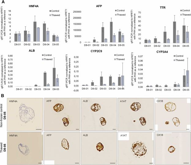
Given that drug-induced liver injury is a leading cause of FDA drug withdrawals, liver models are critical. Using the CERO 3D, researchers generated hepatic organoids using a gene-edited iPSC line, with a nanoluciferase reporter for CYP3A4, enabling them to monitor CYP3A4 expression during differentiation and maturity of hepatocyte-like cells (HLCs). Hepatic organoids were cryopreserved at different stages, and they demonstrated that thawed organoids retained viability and function (Altmaier et al., 2022; Figure 3). This work points toward the creation of organoid biobanks that reduce reliance on donor availability and streamline toxicology workflows.
Figure 3. (A) Validation of non-frozen and day 8 frozen organoids by RT-qPCR with relevant hepatic markers (HNF4A, AFP, TTR, ALB, CYP2C9 and CYP3A4). The organoids were analysed at different differentiation time points from day 8 until day 21 (D8–01: 0 h after thawing, D8–02: 24 h after thawing, D8–03: 3 d after thawing, D8–04: 7 d after thawing, D8–05: 14 d after thawing), mean ± SD for n = 3. (B) Representative ICC figures of non-frozen and day 8 frozen organoids 21 days after thawing with different hepatocyte specific markers, including HNF4A, AFP, ALB, A1AT and CK18. Scale bars = 100 µm.
Advantages of the CERO 3D
- Scalable and Efficient: Cultivate thousands of organoids in a single tube using minimal media.
- Reproducible: Consistent morphology and differentiation across replicates and cell lines.
- User-Friendly: Tubes resemble 50 mL conicals for easy media changes and sampling.
- Flexible: Supports a range of organoid types and can be integrated at any protocol stage.
- Data-Ready: Optional pH monitoring and environmental logging help support GMP workflows.
More Than Just an Instrument
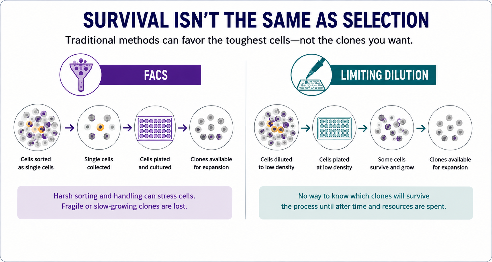
In addition to instrument sales, Cell Microsystems offers lab services for organoid development, allowing researchers to outsource method optimization before transitioning to in-house culture with the CERO 3D. The company also offers CellRaftA microscale polystyrene growth surface within an array used to spatially segregate cells/colonies while maintaining shared media access, enabling imaging over time and targeted isolation. Technology for high-throughput selection and isolation of individual organoids for downstream assays.
References
- Wolfram, et. al. 3R-Compliant Murine Ex Vivo Platform for Scalable Brain Cancer Modeling and Drug Screening. BioRxiv. 2025. doi:10.1101/2025.03.03.641148.
- Fischer, et. al. A complete workflow for the differentiation and the dissociation of hiPSC-derived cardiospheres. Stem Cell Research. 2018; 32:65-72. doi:10.1016/j.scr.2018.08.015.
- Altmaier, et. al. Human iPSC-derived hepatocytes in 2D and 3Dsuspension culture for cryopreservation and in vitro toxicity studies. Rep. Toxicology. 2022; 111:68-80. doi:10.1016/j.reprotox.2022.05.005.

Allysa Stern, Ph.D.
Dr. Stern obtained a Ph.D. and Master of Science in Physiology from North Carolina State University and a Bachelor of Science in Animal Science. Her background is in comparative physiology, with a concentration in cell biology and developing 2D and 3D in vitro models for drug and toxicological studies. Dr. Stern is currently a scientist on the Product Applications team at Cell Microsystems where she focuses on developing novel 2D and 3D cellular workflows using the CellRaft Technology and provides customer training and support for 3D workflows using the CellRaft AIR System.