See these scientific publications for demonstrations of the various uses for products based on the CellRaftA microscale polystyrene growth surface within an array used to spatially segregate cells/colonies while maintaining shared media access, enabling imaging over time and targeted isolation.® Technology.
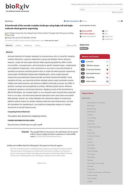
For identifying potential aneuploidy in normal somatic tissues, techniques such as fluorescence in situ hybridization (FISH) and single-cell low-coverage whole genome sequencing (scL-WGS) have been utilized. The accuracy of these techniques including prevalence for false positive/negative accounts of aneuploidy have been inconsistent. To understand the efficiency of these techniques, cells with naturally occurring or induced aneuploidy were generated and isolated with the CellRaft single cell picking system. After CellRaft isolation, these single cells were amplified and sequenced with scL-WGS. The results showed that scL-WGS frequently underestimated aneuploidy levels while FISH overestimated, and a modified 2-probe approach can be used as additional detection for low levels of aneuploidy.
CellRaft Arrays were used as a culture device for seeding gastrointestinal organoidA 3D, self-organizing cell structure, derived from pluripotent or adult stem cells that mimic aspects of organ function and architecture. fragments for studying the effects of microbiota. Using a custom made microinjection system, E. coli was injected into the organoids housed on the CellRaft ArrayConsumables containing thousands of microwells (containing CellRafts) for spatial segregation, imaging, and isolation of single cells, monoclonal colonies, or 3D cultures. and they were imaged to insure proper injection. The Quad Array allowed for 4 different media chambers to test for different conditions and various antibiotics. The use of this customized hardware modification in conjunction with the CellRaft Array allowed for the development of a semi-automated high-throughput organoid microinjection system that could be reproduced in many laboratories.
Gracz AD, Williamson IA, Roche KC, Johnston MJ, Wang F, Wang Y, Attayek PJ, Balowski J, Liu XF, Laurenza RJ, Gaynor LT, Sims CE, Galanko JA, Li L, Allbritton NL, Magness ST
In order to determine the underlying mechanisms of a broad range of issues related to human health and disease, it is important to first understand how somatic stem cells self-renew and differentiate to produce the functional cells of the resident tissue, as stem cells reside in niches where support cells provide signaling critical for tissue renewal. Magness and colleagues demonstrated the use of the CellRaft® Technology to prove that Paneth cells (PC), a known intestinal stem cell (ISC) niche component, enhance organoid formation in a contact-dependent manner. CellRafts® were used to facilitate retrieval of early enteroids for qPCR to correlate functional properties, such as enteroid morphology, with differences in gene expression. This platform enabled the study of a large number of single ISCs simultaneously, either at the clonal level or in the presence of niche cells, with multi-day, three-dimensional culture in extracellular matrices applied directly to the CytoSort™ Arrays. The authors found that direct cell-to-cell contact between ISCs and PCs is required for enhanced ISC growth.
Introduction Regulation of von Willebrand Factor (VWF) activity by a disintegrin and metalloproteinase with a thrombospondin type 1 motif (ADAMTS13) is critical for hemostasis by controlling VWF multimer size. Demonstrating the ADAMTS13-mediated cleavage of VWF under physiological shearFlow conditions intended to resemble those in vivo (e.g., vasculature, catheters, mucosal surfaces). has proven difficult. Traditional analytical methods utilize denaturing environments, thereby poorly replicate the natural vascular environment. This research aimed to develop a sensitive methodology to visualize and quantitate VWF proteolysis by a recombinant ADAMTS13 (rADAMTS13) drug candidate under arterial shear flowFluid movement that applies force to cells and biofilms; used to mimic physiological/pathological conditions. in human blood.
Method A microfluidics-based approach utilizing the BioFlux 1000Z system (Fluxion Biosciences) was optimized by: coating channels with 143 μg/ml collagen type I, blood sample preheating, applying 20 dyne/cm2 pulsatile shear, relying on inherent VWF levels in donor blood, inputting hematocrit based viscosity parameters, and implementing timed protocols. rADAMTS13 was added to healthy donor blood at different concentrations and the time course of platelet adhesion to immobilized collagen was determined by microscopy using fluorescent labeled platelets.
Results Optimized analytical techniques enabled visualization and quantification of VWF proteolysis based on platelet binding under shear flow. Addition of 1.875-7.5 U/ml rADAMTS13 to blood reduced the VWF-mediated platelet adhesion to collagen in a concentration dependent manner. Repeated testing validated the sensitivity. Statistical analysis quantified inter-sample variability.
Conclusion This research successfully established a powerful methodology harnessing microfluidics to gain fundamental insights into rADAMTS13 function under physiologically relevant shear flow conditions. Further enhancement of the techniques, increased biological sampling, and exploration of collagen types could build on these findings.